It is widely known that doing drugs can cause neurological impairments, but it can be difficult to know why. A study published in NeuroImage: Clinical provides evidence that chronic MDMA may not cause white matter lesions that are seen from long-term use of other substances.
MDMA or “ecstasy” is a commonly used illicit drug that causes euphoria. It is frequently consumed at music festivals and in the nightlife scene and is popular due to its ability to increase extraversion, empathy, and sensory perception. MDMA targets serotonin transporters, noradrenaline, and to a lesser extent, dopamine transporters.
Chronic users have been found to display neurocognitive deficits, including difficulty with executive functioning and memory problems, but the underlying mechanisms that cause these deficits is not well understood. Though animal studies have shown axonal degeneration associated with MDMA use, that is unable to be generalized to humans. Human studies on white brain matter in MDMA users have been largely inconclusive, so this new study sought to expand on the body of research.
For their study, Josua Zimmermann and colleagues utilized 39 MDMA users and 39 healthy controls between the ages of 18 and 45-years-old to serve as their sample for this study. Groups were matched by sex, age, education, verbal intelligence, and nicotine use. Exclusion criteria included neurological disease, head injuries, psychiatric disorders other than MDMA abuse, and prior heroin use.
“MDMA users were included if they reported a minimum of 25 lifetime occasions of MDMA use, while having consumed the substance at least once during the 4 months prior to study participation,” the researchers wrote.
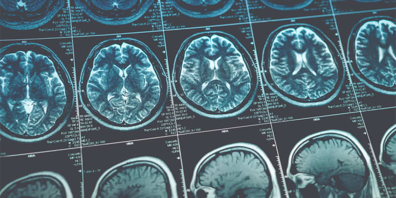
Participants were asked not to utilize illicit substances for 3 days prior to testing and not consume alcohol for 24 hours prior. Participants were screened using a neuropsychiatric interview before receiving an MRI and having blood samples taken. Tractometry imaging was used for the MRI and neurofilament light chain analysis was used for the blood serum samples.
Results showed increased fractional anisotropy, which suggests increased connectivity, for MDMA users in several brain regions, including the internal capsule, corpus callosum, and frontoparietal white matter. However, there was a negative relationship between fractional anisotropy and frequency of usage in the corpus callosum, suggesting the relationships discovered may be dependent on the frequency and dosage of MDMA use.
It is possible that the increase in fractional anisotropy is due to neuronal reorganization due to loss or could be associated with MDMA increasing cerebral blood flow. Future research could explore these possibilities, as it is out of the scope of this study.
This study took important steps into addressing many of the limitations that past research studying white matter in MDMA users has had. Despite this, there are limitations to note. One such limitation is that fractional anisotropy is an indirect measure of white matter structure and can be affected by many factors. Additionally, it is difficult to draw conclusions about the mechanism that is increasing fractional anisotropy.
“We can nevertheless conclude… that – unlike stimulant and ketamine use – chronic MDMA use is not associated with severe white matter lesions but is associated with MDMA-induced changes in white matter diffusion,” the researchers concluded.
The study, “White matter alterations in chronic MDMA use: Evidence from diffusion tensor imaging and neurofilament light chain blood levels“, was authored by