New research sheds light on the effects of spaceflight on astronauts’ brains. The study, published in Scientific Reports, found that going on long space missions and having shorter breaks between missions can cause changes in the fluid inside the brain that may not return to normal before the next spaceflight.
The motivation behind this study was to understand the effects of spaceflight on the human brain, particularly the changes in fluid dynamics and structural morphology. Spaceflight involves various hazards such as radiation exposure, microgravity, and isolation, which can impact the human body in different ways. Previous studies have shown that spaceflight induces changes in the brain, including expansion of the ventricles (cavities filled with cerebrospinal fluid) and shifts in gray and white matter volume.
The researchers wanted to investigate how these brain changes vary based on the duration of space missions and the astronauts’ previous flight experience. They were interested in understanding whether longer missions or shorter recovery periods between missions have a more significant impact on the brain. This knowledge is crucial because as we plan for future long-duration space missions, such as those to Mars, it is important to understand the potential risks and effects on astronauts’ brains.
“Microgravity is a condition in which we have not evolved — it’s fascinating to see how the nervous system changes and adapts in this context,” said study author Rachael Seidler, a professor of applied physiology & kinesiology and professor of neurology at the University of Florida.
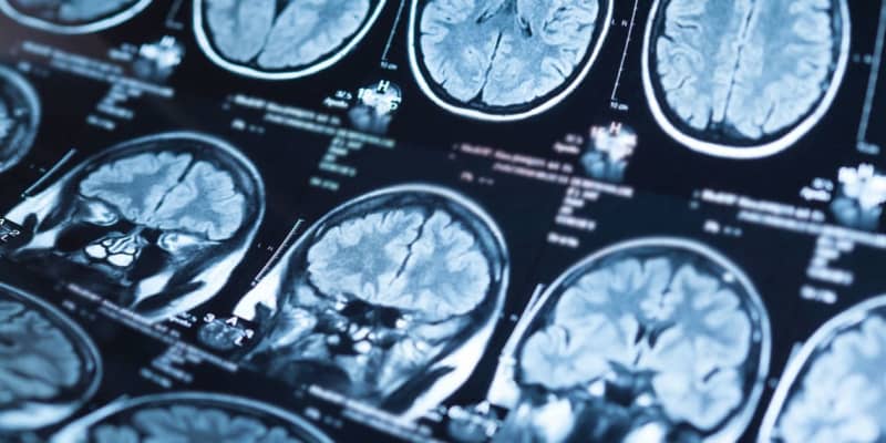
To conduct the study, the researchers scanned the brains of 30 astronauts using magnetic resonance imaging (MRI) before and after their spaceflights. The sample included astronauts who went on two-week missions, six-month missions, and longer missions. They collected T1-weighted anatomical scans and diffusion-weighted MRI (dMRI) scans from the astronauts.
The MRI scans were obtained using a 3 Tesla MRI scanner located at the University of Texas Medical Branch. The T1-weighted images provided structural information about the brain, while the dMRI scans allowed the researchers to analyze white matter microstructure and fluid distribution in the brain.
Seidler and her colleagues found that longer durations of spaceflight led to greater enlargement of certain areas in the brain called ventricles, which are filled with cerebrospinal fluid. However, this enlargement seemed to slow down after about 6 months in space. The study also discovered that astronauts who had more previous flight experience showed smaller ventricle enlargement compared to those who were newer to spaceflight.
“Structural brain changes that occur with spaceflight vary depending on the amount of time spent in space,” Seidler told PsyPost. “Those that travel for a few weeks see little to no changes in ventricular volume — good news for those that are traveling on short space junkets. Those that spent approximately 6 months in space show increased ventricular volume. Those that spent approximately 1 year did not show additional changes, potentially good for even longer planned missions such as to Mars.”
The length of time between successive space missions, known as inter-mission intervals, also played a role in the brain changes. Astronauts who had less than 3 years of recovery time between missions showed little to no ventricular enlargement after their current mission, while those with 3 years or longer of recovery time showed ventricular expansion.
The researchers suggest that when the time between spaceflights is less than three years, it may not be enough time for the ventricles to recover and regain their ability to accommodate the increased fluid in the brain. As a result, the ventricles remain enlarged when astronauts return to space within this shorter time frame.
“I was surprised to see that it may take around three years for the brain to return to preflight after a longer flight,” Seidler said.
There are some limitations to consider in this study. the post-flight MRI scans were done on average about 6 days after the astronauts returned to Earth, with some scans taken as early as 1 day and others as late as 20 days after landing. Although the researchers took into account the time delay between landing and the post-flight MRI scan in their analyses, this timing still could have influenced the observed brain changes.
“We do not yet understand the long term health and human performance consequences of these brain changes,” Seidler added. “I am currently conducting a new study in which we will follow individuals for up to five years post flight.”
The study, “Impacts of spaceflight experience on human brain structure“, was authored by Heather R. McGregor, Kathleen E. Hupfeld, Ofer Pasternak, Nichole E. Beltran, Yiri E. De Dios, Jacob J. Bloomberg, Scott J. Wood, Ajitkumar P. Mulavara, Roy F. Riascos, Patricia A. Reuter‑Lorenz, and Rachael D. Seidler.
