A neuroimaging study of individuals suffering from cocaine use disorder in China showed that these individuals showed signs of atrophy in various brain regions. These alterations were more pronounced in individuals who started using cocaine as minors, before the age of 18. The study was published in theJournal of Psychiatric Research.
Cocaine is a powerful stimulant drug that comes from the coca plant. It is known for its stimulating effects on the central nervous system, leading to increased energy, alertness, and euphoria. However, it is highly addictive and can have serious negative health effects on both physical and mental well-being. Cocaine is considered an illicit drug in most of the world.
In spite of cocaine being illegal, its use is a serious public health problem. There are around 5.5 million cocaine users in the U.S. alone, with nearly a million having cocaine use disorder. Cocaine use disorder is associated with various negative cognitive function and mental health outcomes. These include working memory, attention and executive functioning impairments, abnormal social functioning, decreased quality of life, and also severe cardiovascular disease.
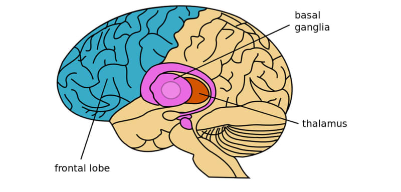
Study author Hui Xu and her colleagues wanted to know whether the structure of the basal ganglia region of the brain might be different in individuals suffering from cocaine use disorder compared to healthy individuals. They also wanted to study if these differences are more pronounced in individuals who started using cocaine at an earlier age.
The basal ganglia are a group of interconnected clusters of neurons located deep within the brain. They are primarily involved in controlling voluntary motor movements, procedural learning, cognition, and in emotional regulation.
The basal ganglia work together to facilitate smooth and coordinated movement patterns. They also play a role in various cognitive functions, including decision-making, habit formation, and action selection, as well as influencing emotional responses and motivation. Dysfunction within the basal ganglia can lead to movement disorders such as Parkinson’s disease and Huntington’s disease, as well as impact cognitive and emotional processes.
The researchers analyzed data from the OpenNEURO database, a free and open database for sharing neuroimaging data. They analyzed records of 68 individuals suffering from cocaine use disorder, who were between 18 and 50 years of age, right-handed and consumed cocaine at least twice a week in the last month before the imaging was done.
They excluded records of individuals who were also dependent on alcohol consumption or tobacco smoking, pregnant or breastfeeding or had some severe systemic disease. The researchers compared these participants to data of 52 healthy individuals used as controls.
The results showed that there were no significant differences between individuals with cocaine use disorder and healthy individuals in regards to overall brain volume or subcortical tissue. However, the researchers identified aberrant surface alterations in subcortical structures in individuals with cocaine use disorder.
Analyses of magnetic resonance imaging recordings showed shrinking and loss of tissue on the surface of the left medial anterior thalamus, right medial posterior thalamus, and right dorsal anterior caudate regions of the basal ganglia.
The magnitude of these changes in the right dorsal anterior caudate was significantly higher in individuals who started using cocaine before the age of 18. The magnitude of changes was lower in individuals who started using cocaine more recently.
“Cocaine use disorder individuals exhibited widespread surface-based alterations of the basal ganglia, including the left medial anterior thalamus, right medial posterior thalamus, and right dorsal anterior caudate, compared to healthy controls, and the surface-based alterations of the right dorsal anterior caudate were significantly associated with the years of cocaine use and onset age of cocaine use,” the researchers concluded.
“These findings shed further light on the effects of cocaine use on the basal ganglia and help us understand the neural basis of cocaine dependence that may provide effective interventions for treating cocaine use disorder.”
The study sheds light on the neural changes affecting cocaine users. However, the study design does not allow any cause-and-effect conclusions to be derived. Additionally, healthy individuals included in this study had a history of tobacco use and some of them also had experience with cocaine use.
The paper, “Cocaine use disorder is associated with widespread surface-based alterations of the basal ganglia”, was authored by Hui Xu, Cheng Xu, and Chenguang Guo.
