In a series of studies on mice, researchers found that the brains of male obese mice don’t signal satiety promptly after eating. Consequently, these mice continue to eat even after being fed, leading to further weight gain. This alteration in response is linked to decreased inhibitory function in the lateral orbitofrontal cortex region of the brain. The study was published in Nature Neuroscience.
Obesity is a medical condition characterized by an excessive accumulation of body fat. It is typically determined by measuring a person’s body mass index (BMI), the weight of the individual divided by the square of height. Generally, individuals with body mass index values of 30 or higher are considered obese.
Obese individuals have a higher risk of various serious health conditions. These include heart disease, type 2 diabetes, certain types of cancer and others. The share of obese individuals has been increasing quickly worldwide in recent decades making obesity a very significant public health problem.
Normally, food intake of mammals, including humans, is regulated by the brain. The classic view of this process was that a deficiency of nutrients would trigger hunger, motivating one to eat. Once satiated, the hunger sensation subsides (satiety). However, studies in recent decades indicated that this process might not be so simple and that it might be disrupted in some individuals, leading to obesity.
Study author Lauren T. Seabrook and her colleagues wanted to test the hypothesis that the mental representations of food and decision-making mechanisms that control food intake are disrupted in obesity. With these mechanisms disrupted, after an individual eats food, he/she would not feel satiated, but would still feel hungry and continue to eat, leading to obesity or further increase in weight.
“We all change how we value food,” said Stephanie Borgland, a professor at the University of Calgary and senior author on the study. “For example, when you’re hungry, a chocolate bar is a high value food. If you were forced to eat five or six chocolate bars though, you would become averse to it. This process is called devaluation.”
Previous studies have indicated that neural structure responsible for this disruption might be located in the lateral orbitofrontal cortex region of the brain. This brain region is known to influence food intake. It is connected to sensory, motor, and limbic brain regions and integrates information from them to guide decision-making.
For the new research, male mice sourced from Charles Rivers Laboratories and the Clara Christie Centre for Mouse Genomics at the University of Calgary, Canada, were used. A genetically distinct breed of mice was also incorporated, enabling researchers to specifically target their inhibitory neurons.
Mice were kept in groups of 3 to 5 per cage. They were divided into two groups. One group was fed a high-fat diet and became obese. This type of obesity is called diet-induced obesity. The other group was fed a normal diet and they stayed lean. Both diets were matched for vitamins, minerals and sucrose. Mice were kept on these diets for a minimum of 12 weeks starting in adulthood.
After this period, researchers conducted a number of trainings and tests on the two groups of mice. In the first experiment, both groups of mice were trained to press a lever to be given a dose of sucrose. However, the number of lever presses needed to get sucrose varied randomly.
After mice learned to press the sucrose release lever, the researchers fed them with the same sucrose solution until they became satiated and then put them in the box with the sucrose release lever. At this point, lean mice showed less goal-directed behavior towards the lever than obese mice. The researchers concluded that the feeling of satiety decreased the value of sucrose for lean mice, but that this did not happen in obese mice.
In the next experiment, these mice were fed until satiety either with sucrose solution or with water, but this was done in the same chamber where the sucrose release lever was. The researchers wanted to test whether the obese mice forget that they were fed or the satiety mechanism is disrupted. Lean mice again showed lower interest in pressing the sucrose release lever compared to obese mice. This showed that the differences in responses was not due to forgetfulness. When these two types of mice are hungry, they press the lever with equal frequencies.
The subsequent experiments showed that lean mice adjust their lever pressing to the expected rewards, but obese mice do not. When the amount of sucrose given was made to be independent of the number of lever presses, lean mice reduced lever pressing, but obese mice did not. Obese mice did not reduce their lever pressing when they were made to feel sick. When both types of mice were switched to the control diet, obese mice remained obese and behaved in a way that indicates that their brains do not decrease the value of food when the mouse is fed (as it normally should).
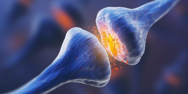
Examinations of the brains of these mice showed that obese mice had reduced inhibition in the neurons of the lateral orbitofrontal cortex. These neurons were more excitable in obese mice. The researchers tracked this to a group of pyramidal neurons in this region of the brain whose inhibition depends on the release of the neurotransmitter gamma-aminobutyric acid or GABA. When the activity of GABA releasing neurons was restored and they started releasing GABA normally in obese mice, the behavior of these mice became similar to that of lean mice.
“Taken together, we causally demonstrate that GABAergic synaptic transmission in the lateral orbitofrontal cortex underlies devaluation [being less motivated to eat by the sight of food in this case] and that increasing GABAergic tone in the lateral orbitofrontal cortex of obese mice restores goal-directed behavior,” the study authors concluded.
“These data propose that obesity-induced changes in lateral orbitofrontal cortex function impede one’s ability to update actions based on current information and suggest that obesity-induced perturbations in lateral orbitofrontal cortex functioning may be an underlying mechanism that contributes to habit-like behavior, which presents an additional challenge for those maintaining food-restrictive diets.”
The study sheds light on the neural mechanisms of food intake regulation in obese mice. However, it should be noted that the study was on mice and not humans. Studies on humans might not yield identical results.
“Our research is confirming that overeating has nothing to do with personal responsibility. It has to do with changes in the way the brain works in response to our food environment,” Borgland said. “There is so much stigma with obesity. You would never hear anyone stigmatising someone with a brain change due to Multiple Sclerosis or Parkinson’s. Why are we doing it with obesity?”
The study, “Disinhibition of the orbitofrontal cortex biases decision-making in obesity”, was authored by Lauren T. Seabrook, Lindsay Naef, Corey Baimel, Allap K. Judge, Tyra Kenney, Madelyn Ellis, Temoor Tayyab, Mataea Armstrong, Min Qiao, Stan B. Floresco, and Stephanie L. Borgland.
