A new study published in Scientific Reports demonstrates striking similarities in synaptic abnormalities and behavioral patterns between male and female mouse models of autism spectrum disorder (ASD). This research challenges the conventional male-centric focus in ASD studies and underscores the vital need to include both sexes in autism research.
Autism spectrum disorder is a developmental disorder characterized by challenges in social interaction, communication, and the presence of repetitive behaviors or restricted interests. The symptoms and severity of ASD can vary widely among individuals, making it a “spectrum” condition, with some needing significant support in daily life while others may be highly skilled or independent. The exact cause of ASD is unknown, but it is believed to be a complex interplay of genetic and environmental factors.
Historically, ASD research has been skewed towards males, prompted by a higher diagnosed prevalence in males compared to females. This new study, however, suggests that the traditional approach may overlook critical aspects of the disorder, especially in females. The motivation behind this research was to bridge this gap and provide a more comprehensive understanding of ASD.
“The main goal is to test the molecular/biochemical differences or similarities between males and females in autism,” said study author Haitham Amal (@haitham_amal), an associate professor at Hebrew University. “The estimated prevalence rate of males diagnosed with ASD exceeds females by a ratio of 4:1. As a result, males remain the primary focus of ASD studies in clinical and experimental settings. Meanwhile, some studies indicate an underestimation of this disorder in females. It was therefore very important to test whether the hypothesis that males are indeed different from females at a molecular and behavioral level.”
The research involved young male and female mice with specific mutations linked to autism. The team compared these mice with regular, non-mutated mice. Two different mouse models were used, each representing a different human-based mutation. The primary objective was to analyze brain connections by examining specific proteins in the brain and the presence of tiny structures in brain cells.
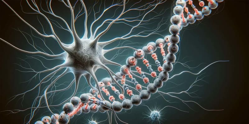
To investigate synaptic development and function, the researchers employed a combination of Golgi staining and the analysis of key neuronal proteins. Golgi staining, a classical neuroanatomical technique, was used to visualize the structure of neurons, particularly the dendritic spines. In parallel, levels of specific proteins critical for synaptic function were measured.
Both male and female mice with ASD-related mutations showed a notable decrease in spine density on their neurons. Spine density is a key indicator of synaptic health and connectivity; lower spine density suggests reduced synaptic connections, which are critical for efficient neural communication.
Alongside this, there were marked reductions in levels of important synaptic proteins, including GAD1, NR1, VGAT, and Syp. These proteins are integral to synaptic transmission and plasticity. GAD1 is involved in the synthesis of the inhibitory neurotransmitter GABA, NR1 is a component of NMDA receptors crucial for synaptic plasticity, VGAT is involved in GABA transport in synaptic vesicles, and Syp is associated with the regulation of neurotransmitter release.
To connect these synaptic changes to behavior, the study employed sociability tests. These tests are designed to assess social interaction and preference for social novelty, which are often affected in individuals with ASD. In these tests, both male and female mice with ASD mutations exhibited deficits in social behavior compared to their non-mutated counterparts. This was evidenced by their reduced interaction with other mice and possibly less exploration of social stimuli.
Importantly, the researchers found a correlation between the observed synaptic changes and the behavioral patterns. The reduced spine density and altered levels of synaptic proteins suggested a disrupted neural network, which could underlie the social interaction deficits observed in the mice. Such a correlation is significant as it provides a potential explanation for the behavioral symptoms of ASD based on underlying neurobiological changes.
“The important and surprising findings are that the biochemical changes and deficiencies in key systems in the brain are the same in both sexes,” Amal told PsyPost. “In addition, we found a decrease in the amount of dendritic spines, which are responsible for the function of the synapse between nerve cells in both sexes. Finally, we examined behavior and found that in the two animal models that each have a different humanic mutation, the social disability appears in both sexes.”
These results challenge the conventional approach in ASD research that has predominantly focused on male subjects, based on the higher diagnosed prevalence of ASD in males. The similarity in synaptic and behavioral patterns found in both male and female mice implies that female models of ASD should not be overlooked, as they provide essential insights into the disorder. This understanding is crucial for developing more comprehensive and effective treatment strategies that cater to both sexes.
However, the study is not without limitations. The use of animal models allowed the researchers to examine the specific biochemical changes associated with ASD. But these models may not fully replicate the complexities of human ASD. Future research could focus on validating these findings in human subjects and exploring the molecular mechanisms underlying these similarities.
The study, “Mutations associated with autism lead to similar synaptic and behavioral alterations in both sexes of male and female mouse brain“, was authored by Manish Kumar Tripathi, Shashank Kumar Ojha, Maryam Kartawy, Igor Khaliulin, Wajeha Hamoudi, and Haitham Amal.
