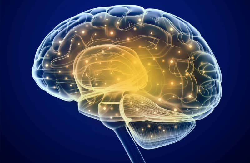
A new neuroimaging study found reduced activation of several regions of the brain that process rewards during a task in which depressed participants expected to be rewarded. These regions include ventral striatum, anterior cingulate cortex/medial prefrontal cortex, anterior cingulate gyrus, angular/middle orbital gyrus, left insula, superior/middle frontal gyrus and precuneus/superior occipital gyrus/cerebellum. The study was published in NeuroImage: Clinical.
The inability to experience pleasure, anhedonia, is one of the key characteristics of depression or major depressive disorder, as it is officially called. It was found to predict poor response to treatment for depression. Anhedonia affects around 37% of individuals suffering from depression and is believed to have a neural basis that has to do with regions of the brain that process our responses to rewards.
In recent years, many studies have tried to identify the brain regions that process our reactions to rewards. Additionally, studies on these neural correlates indicated that the major depressive disorder is also associated with alterations in connectivity between various components of the brain that process rewards and not only in dysfunctions of individual brain areas. However, much of these alterations were yet to be explored.
Aiming to fill this gap in knowledge Hanneke Geugies and her colleagues from the Netherlands devised a study in which they compared activation patterns of areas of the brain that process rewards of patients suffering from major depressive disorder and healthy individuals. Their data came from the Depression in Picture (DIP) neuroimaging study conducted at the University Medical Center Groningen that investigated neural correlates of depression.
Participants were 24 adult patients suffering from at least mild depression as assessed by the Beck Depression Inventory (BDI-II) and the MiniScan. They were compared with a gender-matched group of equal size selected to have no drug or alcohol dependance history, no neurological problems, be fluent in the Dutch language, and have no medical conditions that would prevent magnetic resonance imagining.
Participants underwent magnetic resonance imaging during which they completed 4 sets of cognitive tasks that included monetary rewards.
The tasks “consisted of the presentation of a cue (+€ / ±€ / -€ indicating a reward, neutral or loss trial), a target presentation (blue square), and reward feedback (i.e., +€1.85). Cues and feedback were presented for 1.5s and the target was presented for a fixed duration of 0.5s. Monetary outcomes trials varied for successful reward (+€1.75, +€1.85, +€1.95 and +€2.05) and loss (-€1.60, -€1.70 and -€1.80), but were fixed at +€0.00 for non-reward and neutral trials,” the researchers explained.
Participants received financial compensation for their participation. Rewards won in these cognitive tasks, set to 10 EUR per participant, were added to the total amount of compensation to the participant.
Results showed that the group of depression patients had decreased activation in the ventral striatum part of the brain compared to the healthy group both during the time when reward was anticipated in the cognitive task and when it was received.
The study authors report that “analyses revealed that during reward anticipation, major depressive disorder patients exhibited decreased functional connectivity between the ventral striatum and anterior cingulate cortex / medial prefrontal cortex, anterior cingulate gyrus, angular/middle orbital gyrus, left insula, superior/middle frontal gyrus (SFG/MFG) and precuneus/superior occipital gyrus/cerebellum compared to healthy controls,” the researchers wrote.
Major depressive disorder patients also showed decreased functional connectivity between the ventral tegmental area and left insula regions of the brain compared to healthy controls during the time they were anticipating the monetary reward.
“These results suggest that major depressive disorder is characterized by alterations in reward circuit connectivity rather than isolated activation impairments,” the study authors concluded.
The findings of the study improve scientific understanding of the neural pathways related to symptoms of the major depressive disorder. However, it should be taken into account that the study used just one type of task and that results with different tasks might not be the same. Additionally, 10 of the 24 major depressive disorder patients were receiving antidepressant medication at the time of scanning and this might have had an impact on their brain activity.
The paper, “Decreased Reward Circuit Connectivity During Reward Anticipation in Major Depression”, was authored by Hanneke Geugies, Nynke A. Groenewold, Maaike Meurs, Bennard Doornbos, Jessica de Klerk-Sluis, Philip van Eijndhoven, Annelieke M. Roest, and Henricus G. Ruhé.
